Spinal cord regeneration: translating from salamanders to enhance regenerative repair after injury in mammals
Grant Project Details:
Grant Location
Grant Description
Some salamanders can regenerate a fully functional spinal cord after injury, but humans and other mammals can’t. Dr. Echeverri’s research has identified key molecular differences between salamanders and mammals. She is working to learn more about these differences and how processes in the salamander could be translated to help mammals regenerate spinal tissue.
In response to spinal cord injury humans form dense scar tissue that is inhibitory to the growth of new axons and results in loss of motor and sensory function. To date there is no effective therapy for spinal cord injury or gross neuronal loss as seen in diseases like Parkinson’s or Alzheimer’s. In contrast some lower vertebrates have remarkable regenerative capacity and can functionally regenerate after full spinal cord transection or severe brain injury. Interestingly theseanimals share most genes with humans. We have used comparative approaches to identify which conserved genes are necessary for regeneration in these animals and are now asking what prevents these genes from being activated in humans after spinal cord injury. In addition we have established a 3D in vitro scaffold that allows us to mimic the human spinal cord in vitro, this allows us to image cells directly after injury and to modulate genes directly after injury to ask if activation of these genes can enhance response to injury, this combined approach will hopefully allow us to gradually create a blueprint of pathways that must be activated or suppressed to allow functional regeneration after injury in humans.
In response to spinal cord injury humans form dense scar tissue that is inhibitory to the growth of new axons and results in loss of motor and sensory function. To date there is no effective therapy for spinal cord injury or gross neuronal loss as seen in diseases like Parkinsons or Alzheimers. In contrast some lower vertebrates have remarkable regenerative capacity and can functionally regenerate after full spinal cord transection or severe brain injury. Interestingly these animals share most genes with humans. We have used comparative approaches to identify which conserved genes are necessary for regeneration in these animals and are now asking what prevents these genes from being activated in humans after spinal cord injury. In addition we have established a 3D in vitro scaffold that allows us to mimic the human spinal cord in vitro, this allows us to image cells directly after injury and to modulate genes directly after injury to ask if activation of these genes can enhance response to injury, this combined approach will hopefully allow us to gradually create a blueprint of pathways that must be activated or suppressed to allow functional regeneration after injury in humans.
Grant Awardee Biography

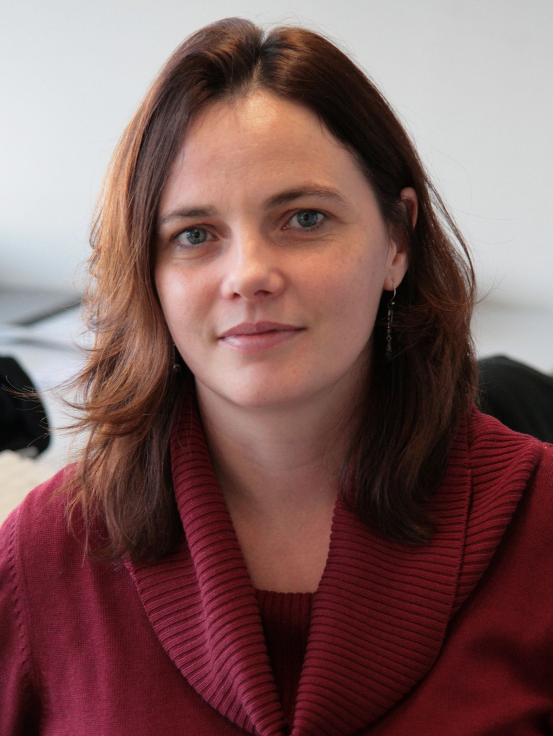
Karen Echeverri, PhD, performed this research as an Assistant Professor in the Department of Genetics, Cell Biology, and Development at the University of Minnesota. She was also a member of the Stem Cell Institute and the Graduate Program in Neuroscience. Dr. Echeverri is currently an Associate Scientist with the Marine Biological Laboratory of The University of Chicago at Woods Hole, MA.


